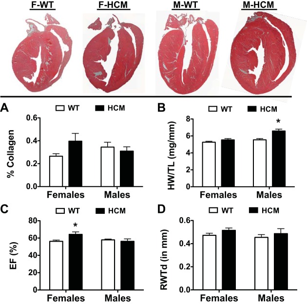
Fig. 4.
Summary of morphometric data from WT and HCM female and male mice. Top, representative hematoxylin and eosin (H&E)-stained longitudinal heart sections from each experimental group. A: bar graph summary of %collagen deposition in each experimental group. Birefringence of collagen fibers was quantified using a semiautomated imaging analysis program. B: HW/TL was determined by dividing cardiac mass (in mg) by tibial length (in mm). Echocardiographic parameters of ejection fraction (EF%; C) and relative wall thickness (RWT; D) calculated from the M-mode measurements. Values are presented as means ± SE. F-WT, n = 6; F-HCM, n = 8; M-WT, n = 8; M-HCM, n = 6. *P < 0.05 from WT counterpart.

