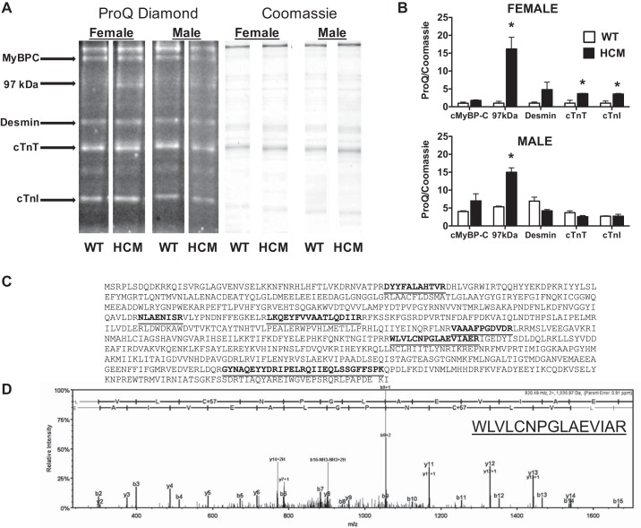
Fig. 6.
ProQ Diamond phosphoprotein stain. A: representative lanes (n = 4/group) cropped from the same gel stained first for phosphorylation (ProQ diamond) and then for total protein (Coomassie). B: bar graph summary of ProQ diamond phosphostain normalized to Coomassie staining in female (top) and male (bottom) mice. Two-way ANOVA identified significant sex and HCM transgene effects and is summarized in text. *P < 0.05 from WT counterpart. C: tryptic digest of 97-kDa band resulted in 6 exclusive peptides and unique spectra highlighted in the peptide sequence of muscle glycogen phosphorylase accounting for 92/842 amino acids (11% coverage). D: LC-MS/MS spectrum of a single fragment with its associated sequence revealed in the inset.

