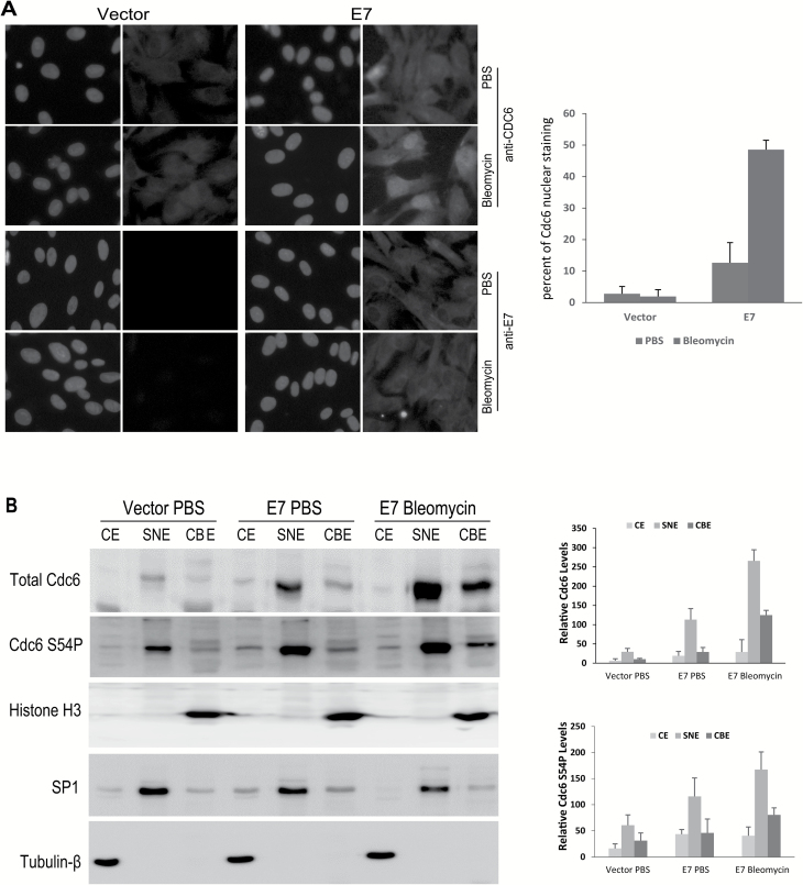
Figure 4.
The localization and subcellular fractionation of Cdc6 in E7-expressing cells. RPE1 cells expressing E7 or vector were treated with bleomycin (3 µg/ml) for 48h. (A) Indirect immunofluorescence microscopy was performed to detect CDC6 and E7 expression and localization (green). 4,5-Diamidino-2-phenylindole dihydrochloride (blue) was used to counterstain the nucleus. Images were captured at ×40 magnification. The data from a representative of at least two independent experiments are shown. (B) The cells were fractionated as cytoplasmic extracts (CEs), soluble nuclear extracts (SNEs) and chromatin-bound fractions (CBEs). The total Cdc6 and Cdc6 S54P levels of each fraction were determined using immunoblotting. β-Tubulin, SP1 and histone H3 were used as loading controls for CE, SNE and CBE, respectively. The data from a representative of three independent experiments are shown in the left panel. The relative levels of total Cdc6 and Cdc6 S54P are summarized in the right panel.

