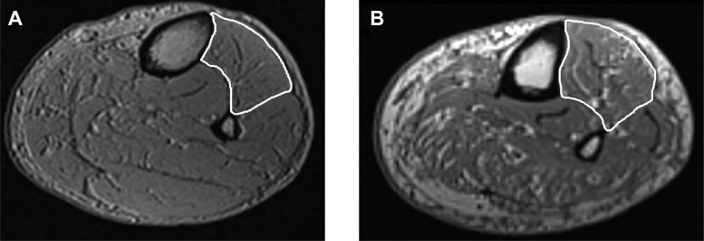
Fig. 2.
Sample MRI images of the leg in an age-matched (∼65 year old) male control (A) and diabetic polyneuropathy (DPN) patient (B; Ref 5). The anterior compartment of the leg is outlined in white. Note: the greater amounts of intramuscular fatty infiltration and noncontractile tissue found in the DPN patient leg compared with the control leg.

