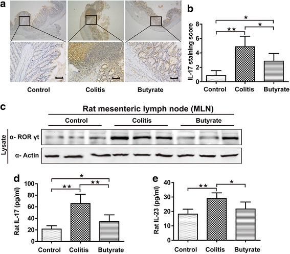
Fig. 4.

Th17 analysis in rats. IL-17 immunohistochemical staining in the colon; upper and lower panel magnifications are × 40 and × 200, respectively. Scale bars, 200 μm (a). Quantified IL-17 immunohistochemical staining in colon (b). Immunoblotting for RORγt in mesenteric lymph nodes, shown are representative western blot results of three rats (c). Plasma IL-17 (d). Plasma IL-23 (e). n = 5–7. Data are the mean ± SE. n = 7. *P < 0.05; **P < 0.01
