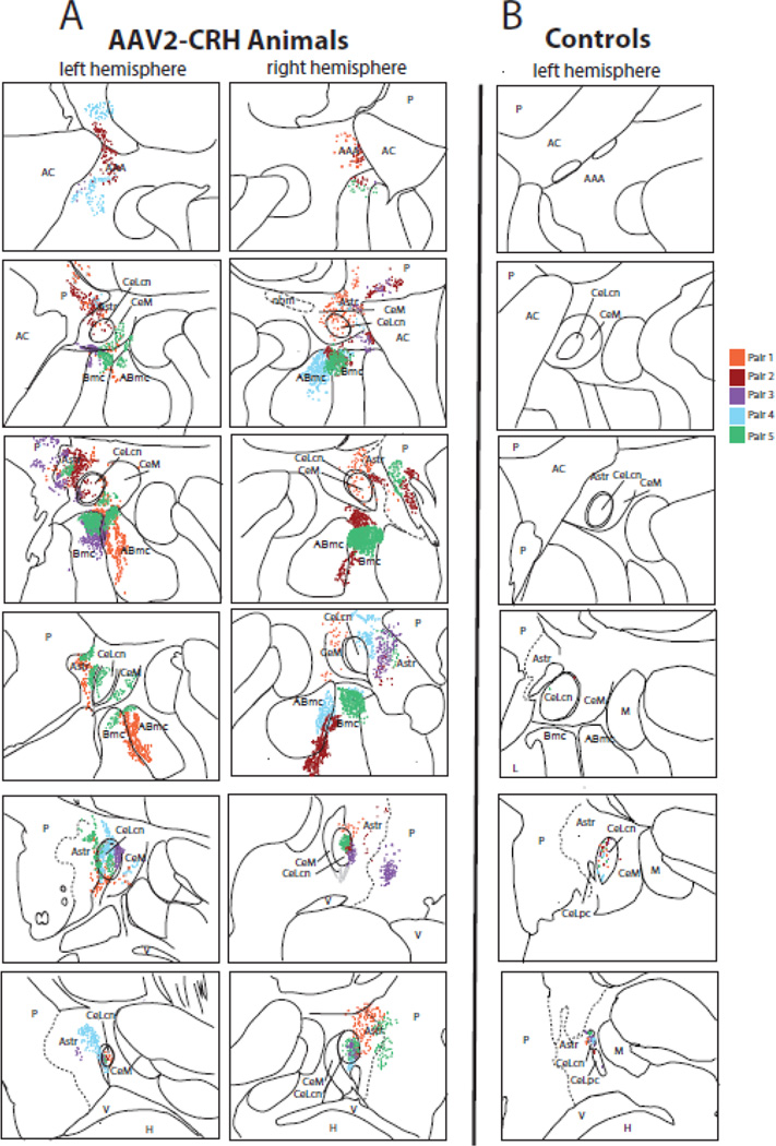
Figure 4. Quantification of dorsal amygdala CRH expression.
(A) Post mortem analyses demonstrated overexpression of CRH in the dorsal amygdala and surrounding regions in the experimental animals, compared to (B) the levels of endogenous CRH observed in the cage-mate control animals (top = anterior, bottom = posterior). Each pair of animals is represented by a different color in the composite image, and each dot represents a CRH expressing cell body. Note that endogenous CRH expression levels in controls were found in the most posterior regions of the Ce (only the left hemisphere is presented), and were substantially lower than that induced by AAV2-CRH transfection. Abbreviations: AAA, anterior amygdaloid area; ABmc, accessory basal nucleus, magnocellular subdivision; AC, anterior commissure; Astr, amygdalostriatal transition zone; Bmc, basal nucleus, magnocellular subdivision; CeLcn, central nucleus, lateral central subdivision; CeLpc, central nucleus, lateral paracapsular subdivision; CeM, central nucleus, medial subdivision; H, hippocampus; L, lateral nucleus; M, medial nucleus; nbm, nucleus basalis of Meynert; P, putamen; V, ventricle.

