
FIGURE 3.
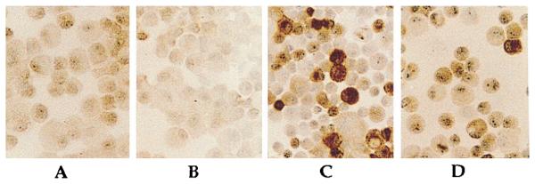
Immunochemical analysis of chondrocytes exhibiting suppression of rhIL-1β-dependent iNOS synthesis by CTS. The iNOS synthesis in chondrocytes was compared in cells subjected to medium alone (A), CTS alone (B), rhIL-1β (C) (1 ng/ml), or costimulation with CTS and IL-1β (D) for 24 h. Histomorphometric analysis of cells showed absence of iNOS in A and B. IL-1β-treated cells (C) show presence of iNOS by intense peroxidase staining in 35 ± 4% of chondrocytes, while cells subjected to simultaneous IL-1β and CTS (D) showed a 62 ± 6% lower staining intensity for iNOS, as compared with iNOS-positive cells in C.