Figure 5.
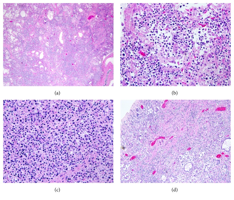
Lymphoma involvement of lung, pancreas, and stomach. (a) Diffuse alveolar and interstitial infiltrates of lymphoma cells in pulmonary parenchyma stained with hematoxylin and eosin (×100). (b) Higher power view of pulmonary infiltrates in a hematoxylin and eosin stained slide (×400). (c) Diffuse infiltrates of lymphoma cells in pancreatic tissue, showing architectural destruction in a hematoxylin and eosin stained slide (×400). (d) Transmural lymphoma cell infiltration in gastric tissue with mucosal erosion in a hematoxylin and eosin stained slide (×400).

