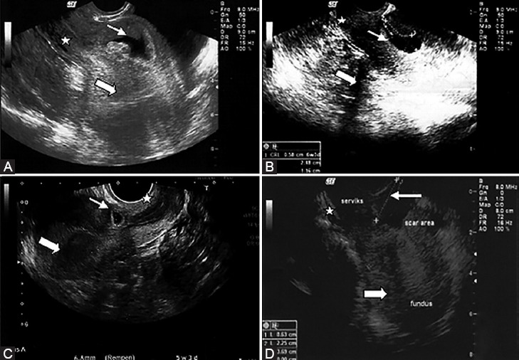
Figure 1.

Transvaginal ultrasonography of the Cesarean scar pregnancy of the 4 women in the study is depicted here. The midline sagittal images demonstrate gestational sacs (small arrows) implanted at the isthmic region between a closed cervix (star) and an empty uterine cavity (large arrows) and the anatomical location of previous Cesarean scars (A-D). A crown-rump length is distinguished inside the gestational sac (A,B).
