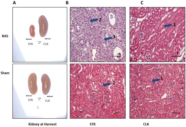
Fig. 2.
A. Representative images of the kidneys of Sham and RAS at harvest. Right cuffed kidney in RAS showing the reduction in the size of right kidney and increase in CLK in RAS. B and C. Representative images of right and contralateral kidney respectively with RAS or sham stained with H&E stain at 200× magnification. Histologic appearance of the sham kidneys (B, C lower) and the contralateral kidney of a mouse with RAS (C, upper) is normal, with back-to-back tubules with abundant eosinophilic (pink staining) cytoplasm and thin, delicate tubular basement membranes (pointed by arrow head 1). The right (stenotic) kidney of a RAS mouse shows severe and generalized tubular atrophy, characterized by a marked reduction in tubular diameter, thickening of tubular basement membranes, and influx of chronic inflammatory cells (pointed by arrow head 2). A relatively presereved tubule is pointed by arrow head 3.

