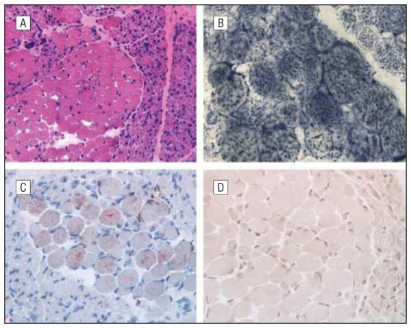
Figure 2.

Muscle biopsy. A, Neurogenic pattern evident in hematoxylin-eosin staining (original magnification ×10). B, Mitochondrial proliferation revealed by succinate dehydrogenase histochemistry (original magnification ×40). C, Some hypertrophic fibers with mild lipid excess demonstrated by oil red 0 staining (original magnification ×20). D, Cytochrome-c oxidase histochemistry showing absence of detectable activity in all fibers (original magnification ×20).
