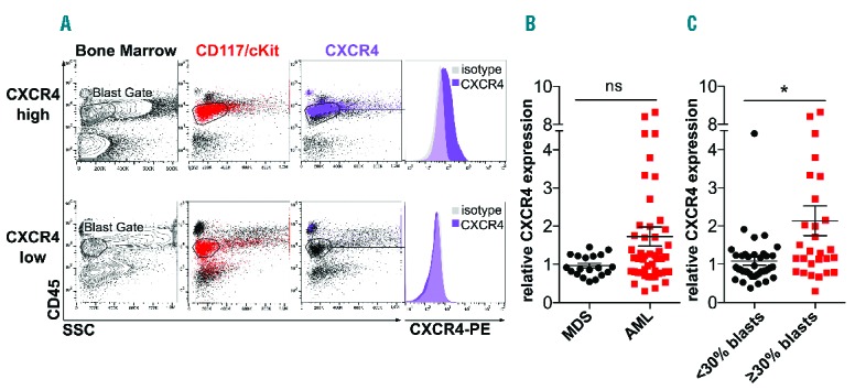
Figure 1.

CXCR4 expression in patients with acute myeloid leukemia (AML) and myelodysplastic syndromes (MDS). (A) Flow cytometric evaluation of CXCR4 surface expression using an anti-CXCR4 antibody. Blasts were gated as CD45low cell population. Anti-CD117 antibody was used for back-gating. Representative data of CXCR4 positive (upper panels) and CXCR4 negative (lower panels) patients are shown. (B and C) Median fluorescence intensity of surface CXCR4 expression relative to isotype control (n=67 patients). Horizontal bars indicate the mean of all individual patient values±SEM; Student’s t-test was used to compare mean relative blast CXCR4 expression. *Statistically significant differences between the groups. (B) MDS versus AML; P=0.062. (C) CXCR4 expression in patients with less than 30% blasts versus CXCR4 expression in patients with at least 30% blasts; P=0.004.
