Figure 1.

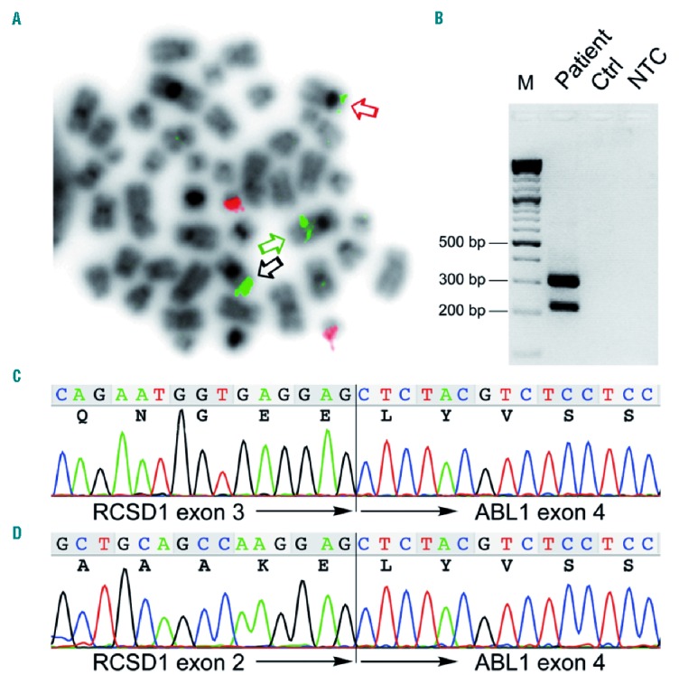
Detection of the RCSD1-ABL1 rearrangement. (A) Fluorescence in situ hybridization (FISH) of a metaphase obtained from the bone marrow at relapse using the BCR/ABL Dual Color, Dual Fusion Translocation Probe (Oncor) showing two signals for BCR (red signals) and three signals for ABL1 (green signals). Black arrow indicates the in situ signal on the normal chromosome 9, the green and the red arrows the ABL1 signals on the der(9) and der(1) chromosomes, respectively. (B) RT-PCR of RCSD1-ABL1 fusion transcripts using primers RCSD1ex1_2-F1 (5′-CCTGAAGGACATGGAGGAAAGACC-3′) spanning exons 1 and 2 of RSCD1 and ABL1ex4-R1 (5′ CTGGATAATGGAGCGTGGTGATG-3′) located in exon 4 of ABL1 showing two distinct amplification products. (C and D) Sequence chromatograms corresponding to the two fusion transcript variants detected by RT-PCR. In-frame fusions of (C) RCSD1 exon 3 to ABL1 exon 4 and (D) alternatively spliced RCSD1-ABL1 lacking RCSD1 exon 3. M: molecular weight marker; Ctrl: control, normal cDNA; NTC: non-template control.
