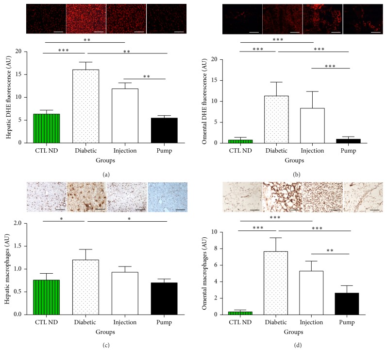
Figure 7.
Reactive oxygen species (ROS) staining in liver (a) and omental tissue (b), macrophage staining in liver (c) and omental tissue (d), and their respective quantifications. The profiles were similar for all quantitative graphs, with diabetic rats having the highest level of oxidative stress or inflammation in both tissue sites. The control nondiabetic (CTL ND) and pump rats had the lowest level of ROS and macrophage staining. The injection group had lower levels than the diabetic group, but higher levels than the CTL ND or pump groups. ∗ p < 0.05, ∗∗ p < 0.03, and ∗∗∗ p < 0.01. Scale bar: 100 μm. DHE, dihydroethidine.

