
Figure 4. Aerobic exercise decreases expression of anti‐lymphangiogenic molecules .
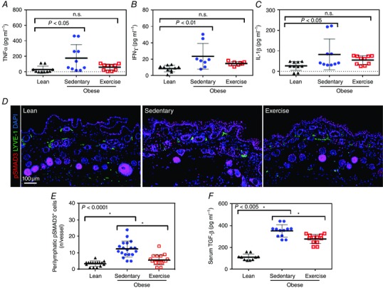
A, enzyme‐linked immunosorbent assay (ELISA) for tumour necrosis factor‐α (TNF‐α) expression in hindlimb tissues from lean, sedentary obese and exercised obese mice (n = 5 or 6 animals per group; one‐way ANOVA: lean vs. sedentary P = 0.0116, lean vs. exercise n.s.). B, ELISA for interferon‐γ (IFN‐γ) expression in hindlimb tissues of mice from various experimental groups (n = 5 or 6 animals per group; one‐way ANOVA: lean vs. sedentary P = 0.0078, lean vs. exercise n.s.). C, ELISA for interleukin‐1β (IL‐1β) expression in hindlimb tissues of mice from various experimental groups (n = 5 or 6 animals per group; one‐way ANOVA: lean vs. sedentary P = 0.0427 and lean vs. exercise n.s.). D, representative photomicrographs of immunofluorescent staining for phosphorylated SMAD3 (pSMAD+; red), lymphatic vessels (LYVE‐1+; green) and nuclei (DAPI; blue) in various experimental groups. E, quantification of perilymphatic pSMAD3+ cells in various experimental groups (n = 5 or 6 animals, with 4 h.p.f. per animal; Student's paired t test: lean vs. sedentary P < 0.0001, sedentary vs. exercise P < 0.0001). F, ELISA for serum transforming growth factor‐β1 (TGF‐β1) in various experimental groups (n = 5 or 6 animals per group; Student's paired t test: lean vs. sedentary P < 0.0001, sedentary vs. exercise P = 0.0015).