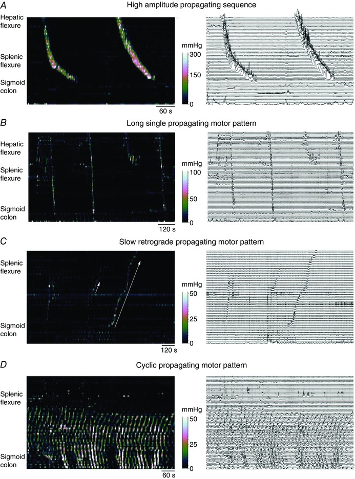
Figure 6. Colonic manometry recordings made using high resolution fibre optic technology taken from healthy adult human colon .

The left‐hand images are displayed as spatio‐temporal colour plots and on the right is the same image displayed as a conventional line plot. A shows the well‐described high amplitude propagating sequences. In B, three long single propagating motor patterns can be seen (black arrows). These motor patterns rapidly propagate across the transverse and descending colon and the component pressure waves have a lower amplitude than the pressure wave shown in A. In C, an example of a slowly propagating retrograde motor pattern is shown (solid white arrow). These originate in the sigmoid colon and over several minutes propagate into the transverse colon. These motor patterns appear during an unstimulated period of recording (i.e. before a meal). Preceding the slow retrograde motor patterns is a short single motor pattern (short white arrow). In D, the cyclic propagating motor pattern is shown. This is the most common postprandial propagating motor pattern. It is largely confined to the distal regions of the colon and propagates predominantly in a retrograde direction, although in this instance both antegrade and retrograde propagation can be seen. The cyclic propagating motor patterns occur at the colonic slow wave frequency of 2–4 cycles min−1. Figure constructed from data published in Dinning et al. (2014 a).
