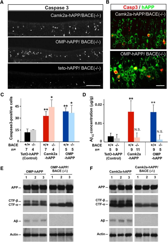
Figure 2.
hAPP-induced apoptosis persisted in a BACE−/− background. A, Cleaved caspase-3 signal in 3-week-old Camk2a-hAPP/BACE−/−, OMP-hAPP/BACE−/−, and control animals. B, The mutants had many more OSNs with caspase-3 signal than controls, which colocalized with the hAPP signal. C, Quantification of caspase-3-positive cells showed that hAPP-expressing lines had significantly more dying cells than the control lines, regardless of BACE genotype (Camk2a-hAPP, 32.8 ± 5.5, n = 7; and OMP-hAPP, 38.5 ± 4.6, n = 5, compared with tetO-hAPP, 11.7 ± 4.3, n = 7; while Camk2a-hAPP/BACE−/−, 44.1 ± 9.2, n = 4, and OMP-hAPP/BACE−/−, 36.3 ± 9.7, n = 5, compared with tetO-hAPP/BACE−/−, 14.8 ± 0.2, n = 4). D, ELISA on OE tissue shows very low levels of Aβ42 concentration in hAPP-expressing mutants with a null BACE background, not significantly different from the levels found in the control lines. E, F, Western blots confirming similar levels of full-length hAPP protein expression in the OEs from OMP-hAPP mice and OMP-hAPP/BACE−/− mice (E), as well as Camk2a-hAPP mice and Camk2a-hAPP/BACE−/− mice (F; individual animals in each lane). Actin was used as a loading control. The expression of α-CTF was also detected across all mutant genotypes, while β-CTF was absent in both the OMP-hAPP/BACE−/− mice and Camk2a-hAPP/BACE−/− mice, consistent with a loss in BACE activity. In addition, Aβ peptide is found in OEs from OMP-hAPP and Camk2a-hAPP mice, but is not detectable in OMP-hAPP/BACE−/− and Camk2a-hAPP/BACE−/− mice, further confirming the lack of BACE activity. All values are reported as the mean ± SD. Scale bars: A, 100 µm; B, 20 µm. *p < 0.01, **p < 0.001.

