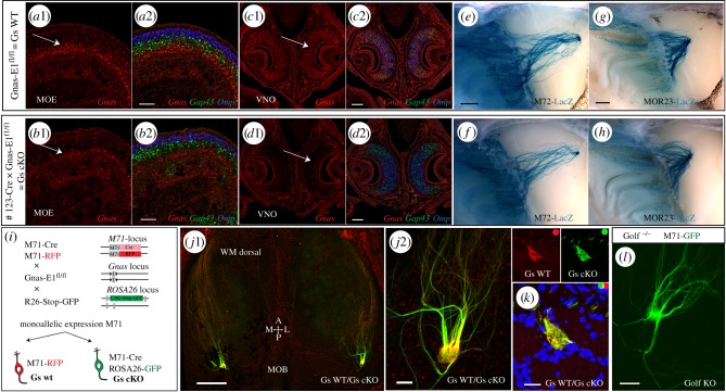
Figure 1.
Conditional deletion of Gs in immature OSNs. (a–d) Three-colour ISH on coronal sections of PD6 MOE. Riboprobes were used against Gnas (red), Gap43 (green) and Omp (blue). (a1) MOE Gnas is expressed more basal than Gap43. (a2) Only a fraction of Gap43 cells colocalize with Gnas. (b1) Gs cKO mice, Gnas expression is no longer observed in Gap43+ OSNs and remaining expression (b2) is more basal. (c1) Gnas is widely expressed in the VNO of Gnas-E1fl/fl mice ( = Gs WT) and (c2) colocalizes with Gap43 and Omp. (d1, d2) VNO of #123-Cre × Gnas-E1fl/fl mice (i.e. Gs cKO mice), Gnas expression is no longer observed. (e, f) Representative images of X-gal-stained medial wholemounts of M72-LacZ OSNs in (e) Gs WT and (f) Gs cKO littermates. Bulbs were analysed for PD10 (Gs WT n = 10; Gs cKO n = 8) and three-week-old (3wo) animals (Gs WT n = 10; Gs cKO n = 6), no mistargeting was observed. (g,h) Representative images of X-gal-stained wholemounts of MOR23-LacZ OSNs in (g) Gs WT (n = 10) and (h) Gs cKO (n = 8) littermates (3wo). No mistargeting was observed. (i) Mice were crossed to obtain animals carrying all four of the indicated targeted alleles (i.e. quadruple mutant). In the quadruple mutant, M71 OSNs are either: (1) RFP+ and Gs WT or (2) GFP+ and Gs cKO. (j1) Wholemount fluorescence of the dorsal bulb in a quadruple mutant described in (i) (6wo). RFP+ Gs WT (red) and GFP+ Gs cKO (green) axons converge and comingle (n > 10 mice); (j2) High magnification view of coalescing axons. (k) Coronal sections of the bulb of a quadruple mutant, showing an M71 glomerulus. Gs WT (red) and Gs cKO (green) axons converge and coalesce. DAPI counterstain. (3wo) (l) Representative wholemount fluorescence image of an M71-GFP glomerulus in a Golf KO mice (2wo). No mistargeting was observed (n = 10 bulbs). MOB, main olfactory bulb; MOE, main olfactory epithelium. Scale bars, 50 μm (a2,b2,j2,l), 100 μm (c2,d2), 500 μm (j1,e,g), 20 μm (k).

