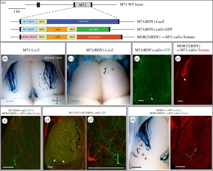
Figure 4.
Signalling-deficient M71(RDY) and MOR23(RDY) ORs induce distinct neuronal identities and differentially regulate anterior-posterior targeting of axons. (a) Schematic overview of the targeted M71 mutations. (b,c) Dorsal view of X-gal-stained wholemounts of M71-LacZ (3wo) and M71(RDY)-LacZ (PD10) mice. (d) Confocal image of a wholemount dorsal bulb from M71(RDY)-caGs-GFP and (e) MOR23(RDY) → M71-caGs-Tomato homozygous mice (PD10). Axons are visualized by intrinsic GFP or Tomato fluorescence. Arrows indicate the main projection sites of lateral axons. (f) Wholemount fluorescence in M71(RDY)-caGs-GFP × MOR23(RDY) → M71-caGs-Tomato mice (PD10). M71(RDY)-caGs-GFP axons (green) project more posterior and do not fasciculate with MOR23(RDY) → M71-caGs-RFP axons (red). (g1, high-magnification view in g2) Wholemount fluorescence in M71-RFP × M71(RDY)-caGs-GFP mice (PD10). M71-RFP (red) and M71(RDY)-caGs-GFP (green) axons project to a similar A-P position. (h) Wholemounts of MOR23 → M71-LacZ × MOR23(RDY) → M71-caGs-Tomato mice (PD10) were X-gal-stained (left) after confocal imaging (right). MOR23 → M71-LacZ (blue, X-gal) and MOR23(RDY) → M71-caGs-Tomato (red, intrinsic fluorescence) axons project to a similar A-P position (pigmentation is used to create anchor points for reference). Scale bars, 500 µm (b,c,h), 250 µm (d,e,f,g1,g2).

