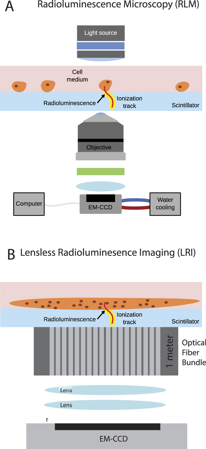
Figure 1. Lensless radioluminescence imaging with an endoscope.

(A) Radioluminescence microscopy (RLM) allows for the high resolution (10 μm) imaging of beta-radiation emitting radiolabeled molecules in individual cells30,31. The advantage of the technique is that decays are converted to an optical signal close to the origin of the decay, which enables decay detection with single-cell resolution. RLM utilizes a thin scintillator plate, which is in contact with the cells of interest, to convert ionizing radiation from emitted beta particles into visible-range photons detectable in a sensitive microscope. (B) On the other hand, lensless radioluminescence imaging (LRI) allows imaging of tissue that is in contact with the scintillator with a much greater field of view but significantly lower resolution because of the lack of an objective lens. The distribution of photons from the radiotracer is directly mapped onto the imaging chip. The compact setup allows for the addition of a fiber bundle endoscope between the sample and scintillator and the detection camera.
