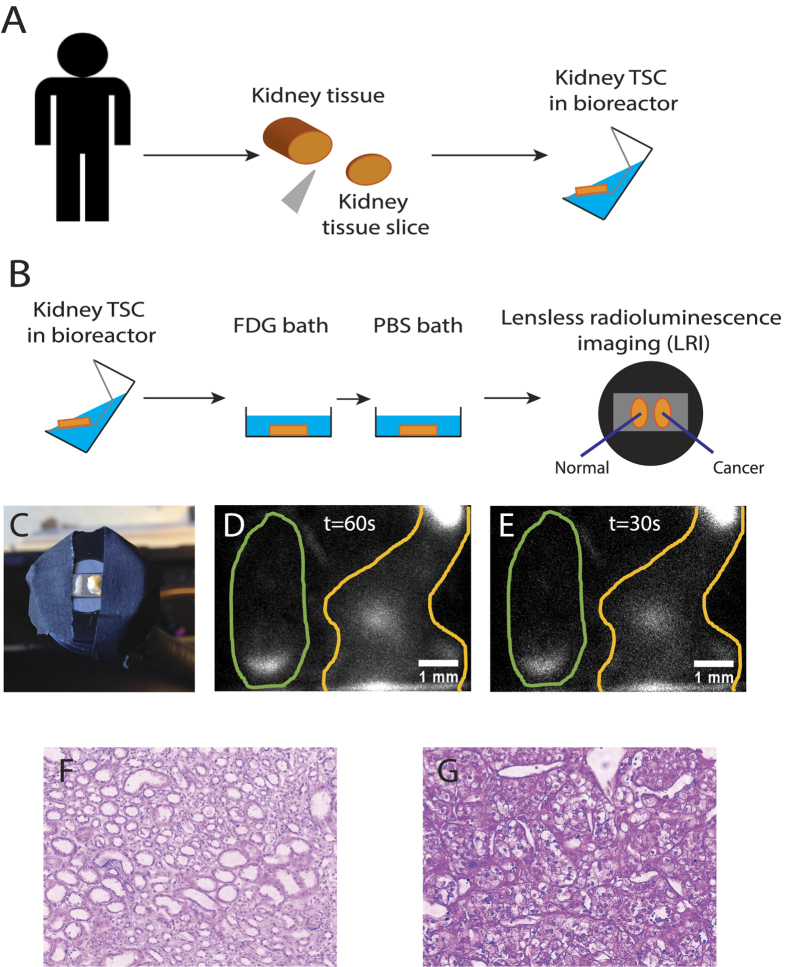
Figure 4. LRI imaging of human tissue slice cultures (TSCs) of renal cell carcinoma and benign kidney tissue.
(A) Surgical removal of cancer tissue and preparation of kidney TSCs. (B) Experimental method for imaging FDG uptake in human renal TSCs. (C) LRI endoscope with a TSCs of benign kidney (left) and RCC (right). (D,E) Images of the distribution of FDG in the TSCs with an acquisition time of 60 s and 30 s, respectively. (F) H&E stain of normal kidney sample. (G) H&E stain of tumor samples shows clear cell renal carcinoma.

