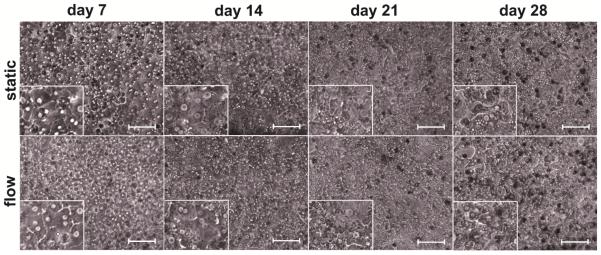
Figure 2.
Representative phase contrast images of primary human hepatocyte co-cultures maintained under static and flow conditions at days 7, 14, 21 and 28. The co-cultured cells showed consistent morphology and nuclear clarity during the first three weeks. Changes in the cell morphology and cytoplasm brining lower nuclear clarity were observed during the fourth week. Image scale bar: 20 μm.

