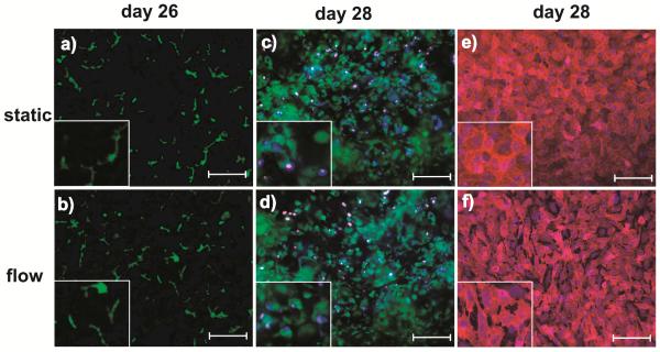
Figure 3. Representative images of hepatic bile canaliculi, EA.hy926 and LX-2 cells in a microfluidic device.
(a,b) CMFDA staining showing the polarization of hepatocytes and formation of a bile canalicular network at day 26. Note that the bile canaliculi were visible in both static and flow conditions. (c,d) LIVE/DEAD staining showing LX-2 cells embedded in the collagen gel of the bottom chamber at day 28 of cell culture. Images were taken ~100 μm below the membrane. Green: live cells, red: dead cells, and blue: nucleus staining with DAPI. (e, f) CD31 staining specific for endothelial cells at day 28. Red: CD31 and blue: nucleus staining with DAPI. Image scale bar: 20 μm.

