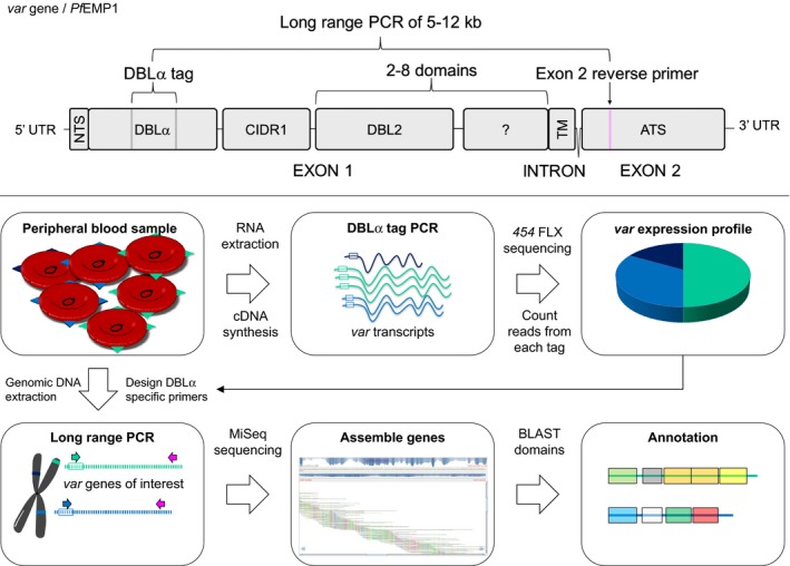
Figure 1. Var gene/PfEMP1 structure and expression analysis work flow.

Var gene structure (upper panel). Var genes consist of two exons, with exon 1 encoding the ectodomain exposed on the infected erythrocyte surface and interacting with human receptors. Exon 1 encodes the N‐terminal segment (NTS), a variable number of DBL and CIDR domains and the transmembrane region (TM). Exon 2 encodes the intra‐erythrocytic part of the protein, the more conserved acidic terminal segment (ATS). Var expression analysis work flow (lower panel). Plasmodium falciparum DNA and RNA was extracted from patient samples. Using cDNA from total RNA, var transcript profiles were generated by counting sequencing reads of PCR‐amplified DBLα sequences using primers targeting semi‐conserved loci flanking the DBLα‐tag sequence. Based on this sequence information, single var gene‐specific 5′ primers were designed for each of the most abundant var transcripts detected by the DBLα‐tag PCR and paired with a 3′ primer targeting a conserved locus in exon 2 to perform a long‐range PCR on genomic DNA. The resulting near full‐length var gene fragments were sequenced, assembled and annotated.
