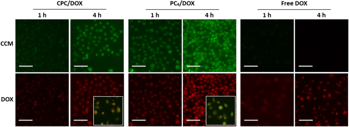
Figure 8. Fluorescence microscope images of PC-3 cells treated with CCM/DOX hybrid nanoassemblies after 1 h and 4 h of treatment.
Green: CCM nanoassemblies, red: doxorubicin. Scale bars indicate 100 μm. The insets represent the merged images of PC-3 cells treated with CCM/DOX hybrid nanoassemblies for 4 h. The yellow signal indicates CCM/DOX hybrid nanoassemblies.

