Figure 2.

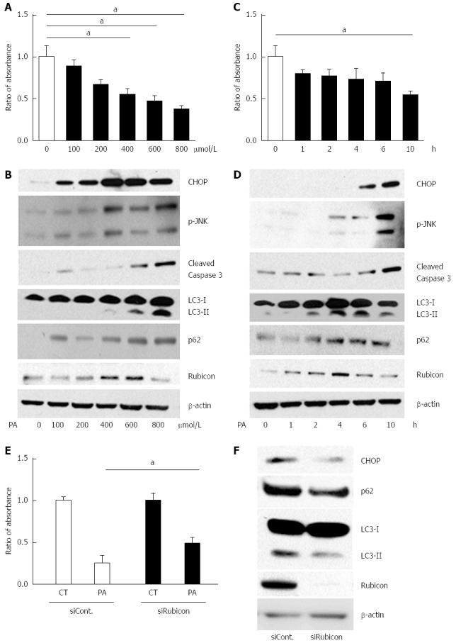
Treatment with palmitate induces apoptosis in a dose- and a time-dependent manner, and initiates but impairs the autophagic process in AML12 cells. A and B: AML12 cells were incubated with PA at the indicated concentrations for 10 h. Untreated AML12 cells were used as the control. A: PA cytotoxicity as evaluated by cell proliferation assay. Living cells are presented as the ratio of absorbance of cells treated with indicated conditions to that of untreated AML12 cells; B: Immunoblotting analyses of CHOP, phosphorylated JNK, cleaved Caspase-3, LC3, p62, Rubicon, and actin. Whole cell lysates were prepared from AML12 cells incubated at the indicated concentrations for 10 h; C and D: AML12 cells were incubated with 800 μmol/L of PA for the indicated periods. Untreated AML12 cells (0 h) were used as the control; C: PA cytotoxicity as evaluated by cell proliferation assay. Living cells are presented as the ratio of absorbance at the indicated incubation time to the absorbance at 0 h; D: Whole cell lysate was prepared from the AML12 cells with 800 μmol/L PA for the indicated incubation times; E and F: AML12 cells were incubated with 800 μmol/L PA for 4 h after transfection with the indicated siRNA: control siRNA (siCont) or Rubicon siRNA (siRubicon); E: PA cytotoxicity as evaluated by cell proliferation assay. Living cells are presented as the ratio of absorbance in AML12 cells with 800 μmol/L PA for 10 h (PA) to that in AML12 control (CT) cells; F: Whole cell lysate was prepared from AML12 cells treated with 800 μmol/L PA with the indicated siRNA. All of the above experiments were repeated three times and representative results are shown. The quantitative data are presented as the mean ± SD; aP < 0.05.
