
Figure 1.
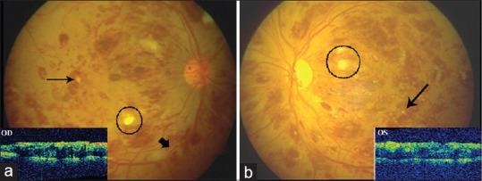
(a) Fundus photograph (OD) showing superficial and deep retinal hemorrhages in all four quadrants with Roth's spots (encircled) in midperiphery, white retinal infiltrates (fine black arrow), and perivascular sheathing (thick arrow). Inset showing spongiform thickening of macula and loss of foveal dip. (b) Fundus photo and optical coherence tomography picture of the left eye (OS) showing similar features