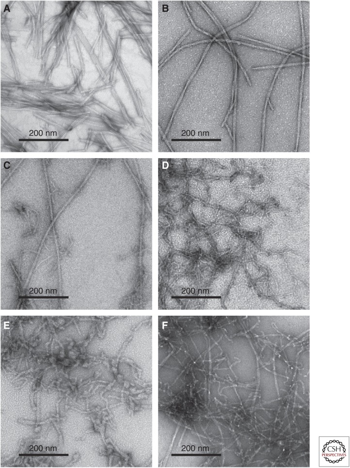
Figure 1.
Negatively stained transmission electron microscope (TEM) images of amyloid-β (Aβ) aggregates. (A) Aβ40 fibrils prepared in vitro with “striated ribbon” morphologies. (B) Aβ40 fibrils prepared in vitro with “twisted” morphologies. (C) Aβ40 fibrils derived from Alzheimer’s disease (AD) brain tissue and prepared by seeding synthetic Aβ40 with amyloid-enriched brain extract. (D) Metastable D23N–Aβ40 protofibrils prepared in vitro. (E) Aβ40 aggregates observed before the appearance of mature fibrils in vitro. A variety of morphologies are seen, including worm-like protofibrils and globular oligomers. (F) Polymorphic Aβ42 aggregates, including both fibrils and protofibrils.

