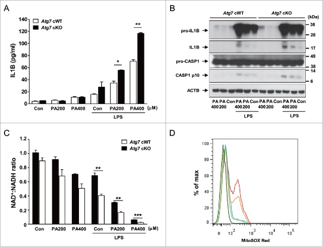
Figure 2.

Inflammasome activation in Mϕs from 12-wk-old mice by varying doses of PA in combination with 500 ng/ml LPS. (A) Primary peritoneal Mϕs were incubated with PA of the indicated concentrations and LPS. After 24 h of culture, IL1B concentration in the supernatant was determined by ELISA. (B) After incubation of primary peritoneal Mϕs with PA of the indicated doses and LPS for 24 h, cell extracts were subjected to western blot analysis using anti-IL1B and -CASP1 Abs. (C) After the same incubation of Mϕs as in (B), NAD+:NADH ratio was determined using a commercial kit. (D) After the same incubation of Mϕs as in (B), cells were incubated with MitoSOX Red for flow cytometry (dark green, unstained; green, Atg7 cWT Mϕs treated with 400 μM PA; blue, Atg7 cWT Mϕs treated with LPS; orange, Atg7 cWT Mϕs treated with 400 μM PA + LPS; red, Atg7 cKO Mϕs treated with 400 μM PA + LPS). *, P < 0.05; **, P < 0.01; ***, P < 0.001.
