
Fig. 1. TPP1 and TPP1-NAbs in CSF after rAAV.caTPP1 infusion.
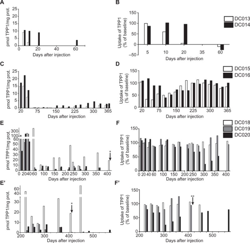
(A, C, E, and E′) An increase in TPP1 in CSF was measured with an enzyme activity assay (see also table S2). (B, D, F, and F′) NAbs in CSF were determined by a neutralization assay: TPP1-deficient mouse embryonic fibroblasts were exposed to a mixture of canine TPP1 plus dog CSF collected before and after treatment. Data were normalized to baseline (pretreatment) values. (A and B) TPP1 activity and NAbs in two dogs (DC013 and DC014) given mycophenolate mofetil treatment starting at day 44 relative to vector infusion (see table S1 for information on vector dose and drug treatment for each dog). (C and D) TPP1 activity and NAbs in two dogs (DC015 and DC016) given mycophenolate mofetil starting at day 33 relative to vector infusion. (E, E′, F, and F′) TPP1 activity and NAbs in three dogs (DC018, DC019, and DC020) given mycophenolate mofetil starting at day −5 relative to vector infusion. Vector infusions were at about 90 days of age in all cases. *TPP1 in CSF from DC019 was 0.3 pmol/mg protein or 100% of normal. **CSF from DC020 showed no detectable TPP1 uptake at this time point, indicating high levels of NAbs.