Fig. 3.
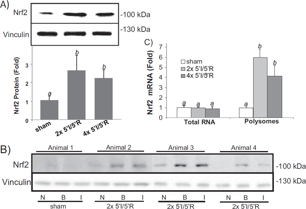
Brief cycles of I/R cause elevation of Nrf2 protein. Mouse hearts were subjected to 2× or 4× 5′I/5′R. Tissue extracts from the whole hearts were used for Western blots (80 µg protein/lane) with vinculin as a loading control (A). The bar graph represents the relative density of the Nrf2 band over vinculin in the same sample as means ± standard deviations from 13 animals (A). Ischemic (I) versus non-ischemic (N) areas and 1–2 mm border zone (B) were rapidly dissected following Evan's blue dye perfusion for Western blot (B). A sham operated control was used for collecting areas corresponding to I, N or B (B). Total RNA or polysomal RNA was extracted from the whole heart tissue lysates for real time RT-PCR (C). The data represent means ± standard deviations from 3 (total RNA) or 4 animals (polysomal RNA, C). ANOVA was used to determine the significant difference (p < 0.05) between the means. The means labeled “a” are significantly different from that labeled “b”.

