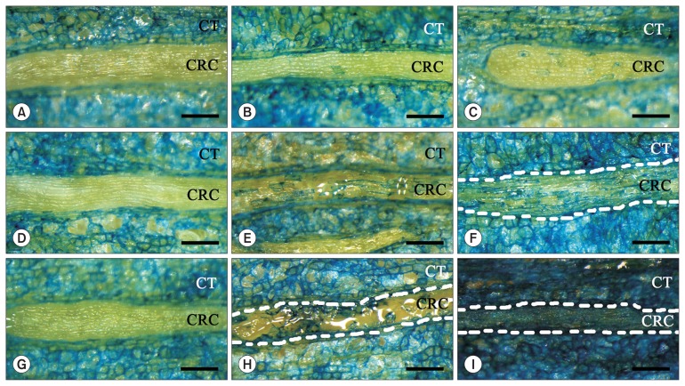
Fig. 2.
Evans blue-stained dead cells in the epithelium of the cortical resin canals in current-year main stem cuttings of 5-year-old Pinus thunbergii. Abbreviations represent cortical resin canals (CRC) and cortical tissues (CT). Scale bars = 0.25 mm. (A, D, G) Stem cuttings inoculated with distilled water shown 1, 3, and 7 days after inoculation (DAI), respectively, and no dead cells were found in the cortical resin canal. Data from the article of Son et al. (2014) (Nematology 16:663–668) with permission, conducted at the same time as the experiment in this study. (B, C) Stem cuttings inoculated with Bursaphelenchus mucronatus, 1 DAI. Note that blue-stained dead cells are sporadically distributed in the resin canal; the area covered by the blue-stained dead cells was 5.53% (C). (E, F) Stem cuttings inoculated with B. mucronatus, 3 DAI. The areas covered by the blue-stained dead cells were 19.53% and 30.18%, respectively. The resin canal is present between the 2 dotted white-lines (F). (H, I) Stem cuttings inoculated with B. mucronatus, 7 DAI. Enlarged epithelial cells are observed between the 2 dotted white-lines (H). The resin canal is also present between the 2 dotted white-lines. All the epithelial cells in the resin canal were dead (I).

