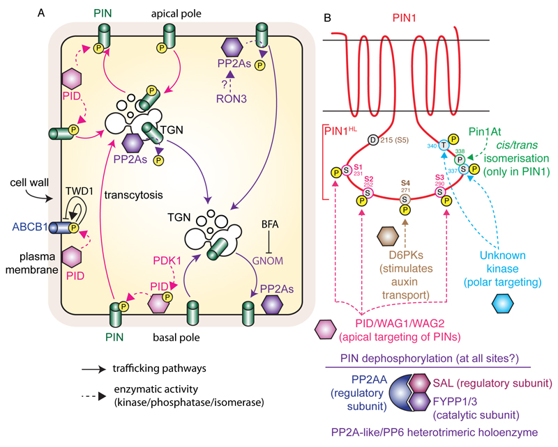
Figure 2. Model for PID and PP2As antagonistic activity on PIN phosphorylation, and the dual action of ABCB1 phosphorylation by PID.
A) Non-phosphorylated PINs undergo continuous endocytic recycling in a GNOM-dependent manner, which mediates their basal localization. The PID kinase phosphorylates PINs at the plasma membrane, which prohibits them from recycling via the GNOM-dependent route. Phosphorylated PINs instead undergo transcytosis to the apical pole of the cell. PINs are dephosphorylated by PP2As phosphatases, presumably at the plasma membrane and endosomes. PID also phosphorylates ABCB1, which promote ABCB1-mediated auxin export in the absence of TWD1, but inhibits it in its presence. B) Schematic representation of PIN1 topology and position of PIN1 phosphorylation site within PIN1 hydrophilic loop (PIN1HL). The action of the kinases/phosphatases/isomerase on specific residues is highlighted. Note that only D6PK main phosphorylation sites (S4 and S5) are highlighted, but that S1, S2 and S3 are also phosphorylated by this kinase, albeit less potently. Similarly, PID/WAG1/WAG2 are able to phosphorylate S4 and S5 but preferentially act on S1, S2 and S3. Dashed arrows represent direct phosphorylation, dephosphorylation or isomerisation events; arrows with chevron-shaped arrowhead represent trafficking pathways and blunt-ended lines represent inhibition. Yellow-filled circles with the letter ‘P’ represent phosphorylation, while grey-filled circles represent individual amino acid (each residue is numbered according to its position within the PIN1 protein).

