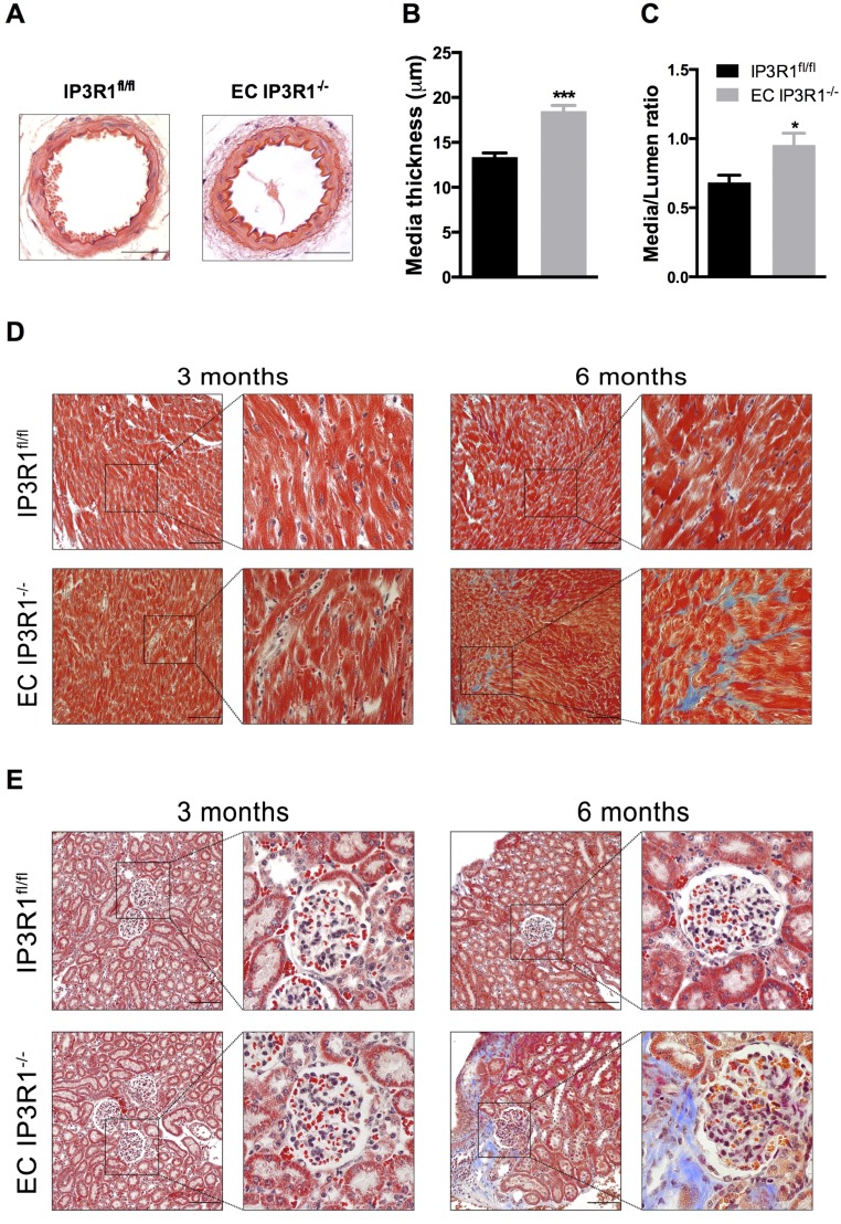
Fig. S4.
Targeted end-organ damage in EC IP3R1−/− mice. (A) Representative pictures of H&E staining on paraffin-embedded mesenteric arteries of 6-mo-old IP3R1fl/fl and EC IP3R1−/− mice. Data statistics of media thickness (B) and media/lumen ratio (C) calculated from at least eight vessels of four individual mice per each group are shown. *P < 0.05; ***P < 0.001 vs. IP3R1fl/fl mice analyzed by Student’s t test. Masson’s trichrome staining on paraffin-embedded sections shows massive fibrosis (indicated by blue stain) of both the heart (D) and kidney (E) in 6-mo-old EC IP3R1−/− mice, which is hardly detected in IP3R1fl/fl littermates. (Scale bars: 50 μm.)

