Fig. 1.
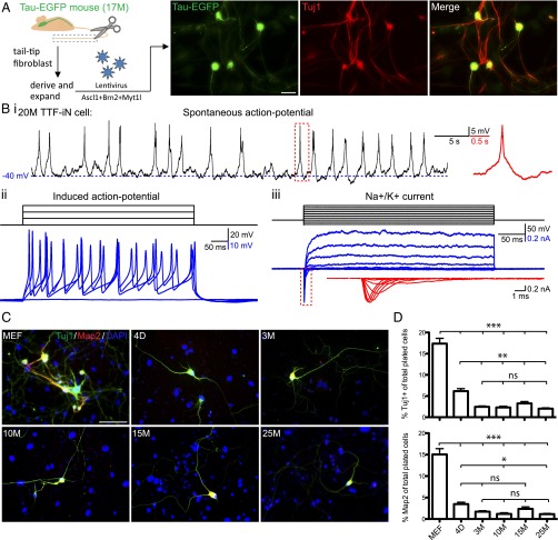
Fibroblasts from postnatal and adult mice are less susceptible to iN cell reprogramming than MEFs. (A) Experimental approach (Left) and image of iN cells (Right) generated from TTFs of a 17-mo-old (17M) Tau-EGFP animal. Micrograph showing Tau-EGFP epifluorescence (green) and immunofluorescence detection with Tuj1 antibodies (red). (Scale bar, 30 μm.) (B) Sample traces of spontaneous AP firing (i), current-pulse–induced AP (ii), and voltage-gated Na+ and K+ currents (iii) recorded from TTF iN cells reprogrammed from a 20-mo-old animal (20M TTF-iN cell). Insets in red, magnified view of corresponding boxed area. (C) Representative images (Left) for Tuj1 (green) and Map2 (red) immunoreactivity of TTF iN cells generated from embryonic (MEF), postnatal (4 d old; 4D), adult (3 mo old; 3M), middle aged (10 and 15 mo old; 10M and 15M, respectively) and aged (25 mo old; 25M) animals, 3 wk after induction. (Scale bar, 50 μm.) (D) Average fractions of Tuj1+ (Top Right) and Map2+ (Bottom Right) cells 3 wk after infection of initially plated cells. Data are presented as means ± SEM (n = 3–5 experiments with three technical replicates each). Significance was determined by using one-way ANOVA with Bonferroni post hoc test (*P < 0.05; **P < 0.01; ***P < 0.005; ns, not significant).

