Fig. 3.
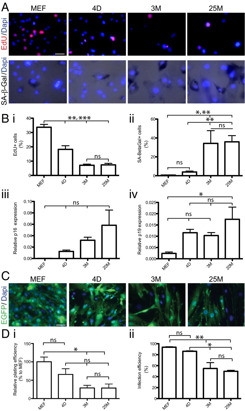
Aging-associated features in donor fibroblasts. (A) Representative images indicating age-dependent decrease of EdU incorporation (EdU staining in red, Top) and increase in senescence associated β-Gal activity (black staining, Bottom) in fibroblasts from different age groups (MEFs, 4D, 3M, and 25M, Left to Right) counterstained with DAPI (blue). (B) Average bar graphs indicate means ± SEM of percentages of EdU+ (i), SA-β-Gal+ (ii) fibroblasts and average relative mRNA levels for senescence markers p16 (iii) and p19 (iv) measured by qRT-PCR. Asterisks indicate significant difference (n = 3 independent batches; *P < 0.05; **P < 0.01; ***P < 0.005; ns, not significant; ANOVA with Bonferroni post hoc test). (C) Representative images showing lentivirus-mediated EGFP expression (green) and DAPI (blue) staining in MEF, 4D, 3M, and 25M TTF. (D) Average plating efficiency (DAPI staining/field of view, normalized to MEF condition; i) and infection efficieny (percentage of EGFP+/DAPI-stained cells; ii) calculated for MEF, 4D, 3M, and 25M TTF (left to right). Bar graphs represent mean ± SEM, and statistical significance were calculated by using ANOVA with Bonferroni post hoc test (n = 3 batches, *P < 0.05, **P < 0.01; ns, not significant). (Scale bars, 20 μm.)

