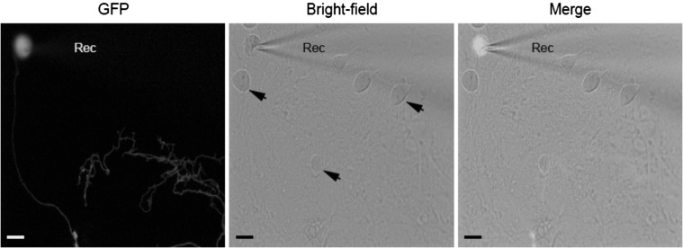
Fig. S2.
Patch-clamp configuration for postsynaptic recording (related to Fig. 2). TTF-derived iN cells were additionally infected with lentivirus expressing GFP and cocultured with low-density mouse primary hippocampal neurons for 3 wk to allow them to form synaptic connections. Left, GFP fluorescence view; Center, bright-field; and Right, as both views merged. Rec, recording electrode, placed on a GFP+ TTF-iN cell. Black arrowheads (Center), non-GFP primary neurons. (Scale bars, 10 μm.)

