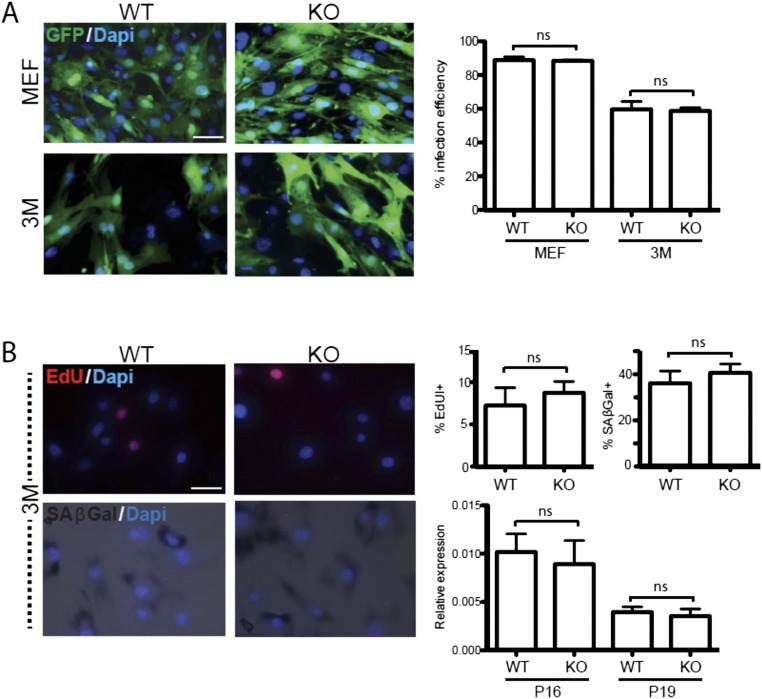
Fig. S5.
Germ-line loss of FoxO3 does not affect infection efficiency, proliferation, or senescence (related to Fig. 4). (A) Representative images (Left) of infection efficiency (as quantified with lentivirus expressing GFP, 24 h postinduction) for TTFs derived from embryonic (MEF; Upper) or 3-mo-old (3M; Lower) WT (Left) and FoxO3−/− (KO, Right) littermate animals. Average percentages of GFP-expressing cells (Right) were calculated with respected to total DAPI staining (blue). (B) Sample images of 3M TTFs stained with EdU (Upper Left) and SA-β-Gal (Lower Left), respectively, used as proliferation and senescence markers. Average quantifications for EdU/SA-β-Gal staining (Upper Right), as well as p16/p19 mRNA expression levels (as measured by qRT-PCR; Lower Right) for WT and KO conditions are indicated. For all graphs, average values represent means ± SEM, n = 3 independent batches. No significant differences were detected in either experimental conditions (ns, P < 0.05, ANOVA with Bonferroni post hoc test) for either parameter tested. (Scale bars, 20 μm.)

