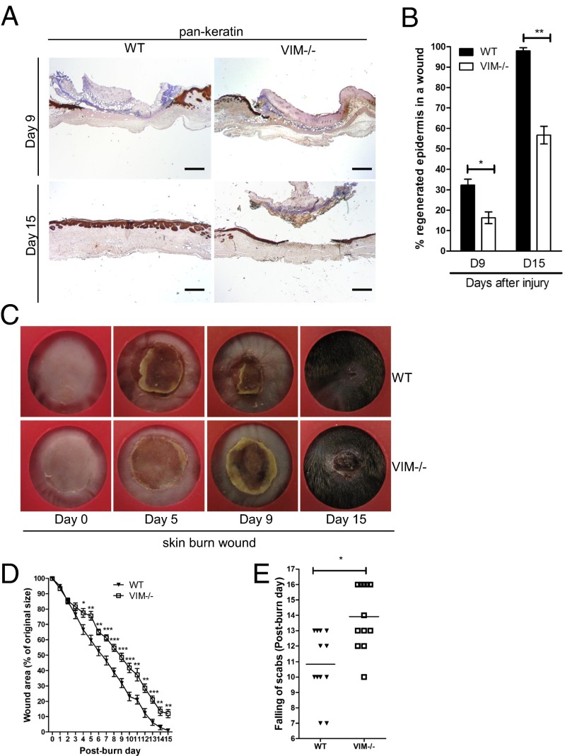
Fig. 1.
VIM−/− mice display wound-healing defect in a burn wound model. (A) Representative pictures showing immunohistochemical labeling of pan-keratin in WT and VIM−/− wounds on days 9 (D9) and 15 (D15) postinjury. (Scale bar, 100 μm.) (B) Quantification of the percentage of wound reepithelialization at different time points after wounding in VIM−/− and WT wounds. Data are shown as means ± SEM; n = 6. (C) Representative wound pictures from VIM−/− and WT mice during the 15-d wound-healing period. (D) Quantification of the remaining wound area at different time points after wounding in WT and VIM−/− groups. Data are shown as means ± SEM; n = 6–12. (E) Comparison of the healing times (scab falling off) in the days after wounding. Data are shown as means ± SEM; n = 12. *P < 0.05; **P < 0.01; ***P < 0.001.

