Fig. 2.
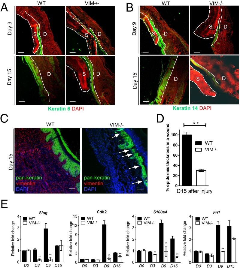
Compromised reepithelialization and EMT differentiation in VIM−/− wounds. (A and B) Representative pictures of confocal images of keratin 6 (green) and DAPI (red) (A) and keratin 14 (green) and DAPI (red) (B) in VIM−/− and WT wounds on day 9 and 15 after skin burn injury. D, dermis region; S, scab region. (Scale bars, 200 μm.) (C) Representative confocal images of keratin expression as visualized by a pan-keratin antibody (green), vimentin (red), and DAPI (blue) in VIM−/− and WT wounds on day 15 postinjury. White arrows indicate the region of thin and poor keratinization in VIM−/− wounds. (D) Quantification of average epidermis thickness on day 15 after burn injury. Bars indicate the mean fold changes relative to WT ± SEM; n = 3. (E) qRT-PCR analysis of mRNA transcripts for Slug, N-cadherin (Cdh2), FSP-1 (S100a4), and fibronectin (Fn1) in isolated epidermal regions of VIM−/− and WT wounds on days 0, 3, 9, and 15 after burn injury. Bars indicate the mean fold changes ± SEM relative to day 0 WT; **P < 0.01; n = 3.

