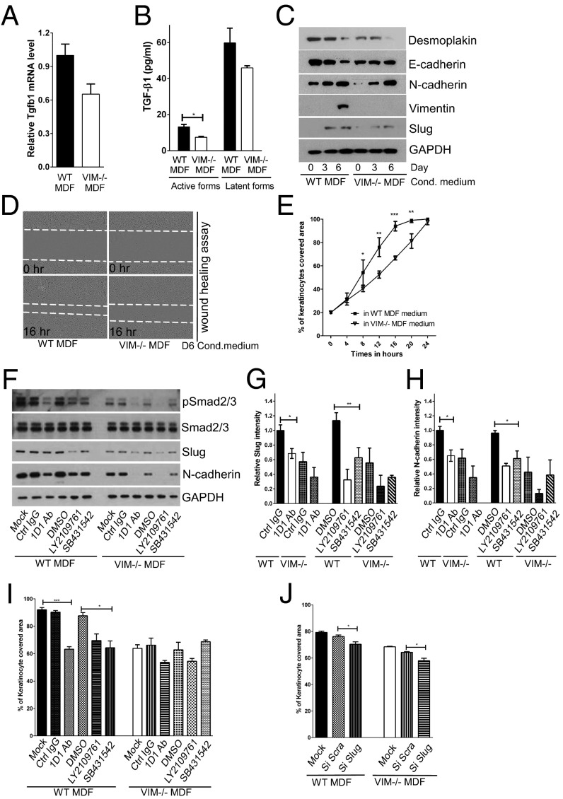
Fig. 4.
Vimentin promotes TGF-β production from fibroblasts driving EMT and migration of keratinocytes. (A) qRT-PCR analysis of transcripts for TGF-β1 (Tgfb1) in VIM−/− and WT MDFs. Bars show mean fold changes ± SEM relative to WT; n = 6. (B) Level of active and latent forms of TGF-β1 in the supernatants of 6-d MDF cell cultures were analyzed by ELISA. Data are shown as mean ± SEM; n = 3. (C) VIM−/− and WT MDF cell-culture media were extracted on 0, 3, and 6 d after cell growth. The growth medium of mouse keratinocytes was replaced with the MDF-conditioned medium for 5 d. Cell lysates were collected and blotted with antibodies against desmoplakin, E-cadherin, N-cadherin, vimentin, Slug, and loading control GAPDH. (D and E) In vitro wound-healing assay of mouse keratinocytes grown in 6-d conditioned medium from VIM−/− MDFs and WT MDFs. The cell gap was monitored over 24 h, and the wound areas were measured and plotted against the time point. At least four wound scratches were analyzed per experiment. Data are shown as means ± SEM; n = 3. (F) Mouse keratinocytes grown in 6-d conditioned medium from the VIM−/− and WT MDFs were treated with control IgG (Ctrl IgG, 10 μg/mL), pan–TGF-β neutralizing antibody 1D11 (1D1 Ab, 10 μg/mL), two chemical inhibitors of TGF-β receptors [LY2109761 (2 μM) and SB431542 (2 μM)] or were grown in the corresponding DMSO control (DMSO, 2 μM) for 3 d. The cell lysates from this experiment were blotted with antibodies against pSmad2/3, total Smad2/3, Slug, N-cadherin, and loading control GAPDH. (G and H) Quantification of Slug and N-cadherin intensity in F equalized to GAPDH; bars show the mean fold changes relative to WT Ctrl IgG ± SEM; n = 3. (I) In vitro wound-healing assay of mouse keratinocytes (at 16 h of wound healing) in the treatments in F. (J) The y axis shows the percentage of area covered (at 16 h of wound healing) by keratinocytes transfected with mock, scramble siRNA (si Scra), or Slug siRNA (si Slug) oligos for 2 d and incubated in 6-d WT or VIM−/− MDF-conditioned medium. In I and J, at least four wound scratches were analyzed per experiment. Data are shown as means ± SEM; n = 3; *P < 0.05; **P < 0.01; ***P < 0.001.

