Fig. 5.
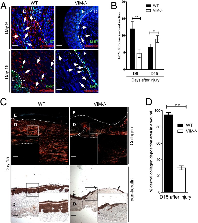
Vimentin promotes mesenchymal cell proliferation and collagen accumulation in vivo. (A) Representative confocal images of the expression of Ki67 (punctate green signal in the nucleus) and vimentin (red) in VIM−/− and WT wounds on days 9 (D9) and 15 (D15) after burn wounding. Nuclei were counterstained with DAPI (blue). (Scale bars, 20 μm.) The white arrowheads indicate examples of ki67+ cells in dermal regions. D, dermis region; E, epidermis region. (B) Quantitation of ki67+ cells in mesenchymal/dermal regions of wounds. (C) Representative pictures of Picro-Sirius Red staining of collagen (Upper) and the immunohistochemical labeling of pan-keratin (Lower) in the corresponding sections of VIM−/− and WT wounds on day 15 postinjury. The right lower corner of each panel shows an enlarged image of the area in the white box. (Scale bars, 100 μm.) (D) The quantitation of collagen accumulation (Picro-Sirius Red-positive areas) in mesenchymal/dermal regions of wounds. In B and D data are shown as means ± SEM; n = 3; *P < 0.05; **P < 0.01.

