Fig. S1.
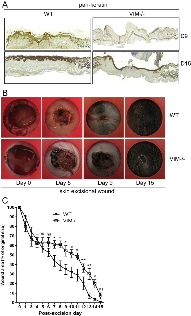
VIM−/− mice have slower wound healing in an excisional wound model (related to Fig. 1). (A) Representative pictures of immunohistochemical labeling of pan-keratin in wounds in VIM−/− and WT mice on days 9 (D9) and 15 (D15) postinjury. (B) Representative pictures of wounds in WT and VIM−/− mice 15 d after excisional injury. (C) Quantification of the wound area remaining at different time points after wounding in the WT and VIM−/− groups. Data are shown as means ± SEM; n = 4. *P < 0.05; **P < 0.01; ns, not significant.

