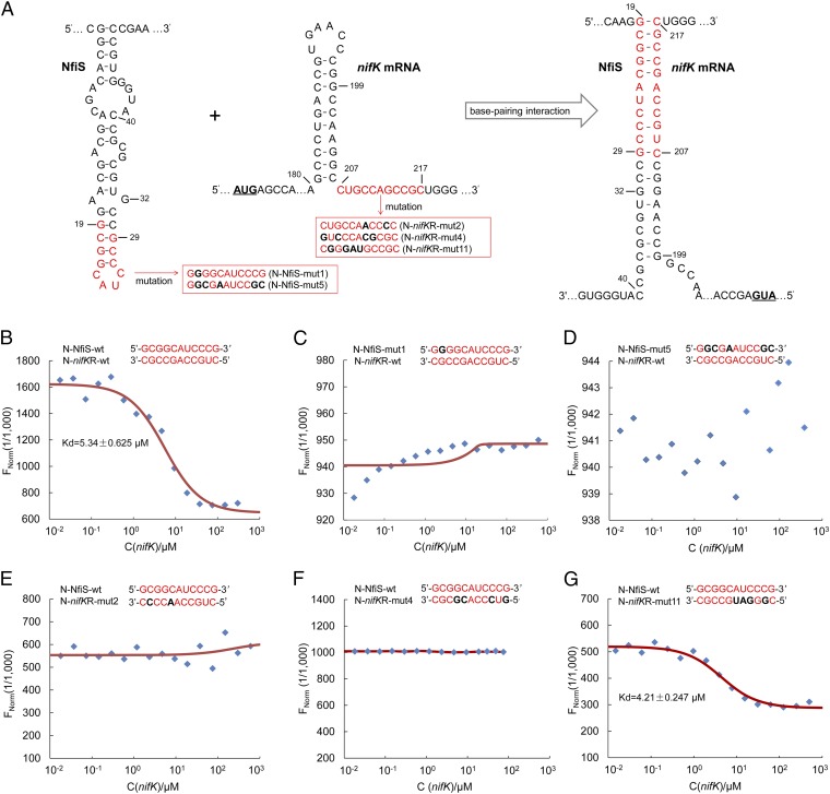
Fig. 3.
Binding of NfiS to nifK mRNA. (A) Schematic representation of the base-pairing complex formation (Right) between the NfiS stem loop (Left) and the complementary sequence of nifK mRNA (Middle). Pairing nucleotides are shown in red. Point mutations introduced into synthesized oligonucleotide derivatives in red boxes are shown in black and bold. mut, mutation; N, synthesized oligonucleotide; nifKR, nifK mRNA; wt, wild type. (B–G) Determination of the binding affinity of NfiS to nifK mRNA by microscale thermophoresis. The concentration of labeled N-NfiS molecules was constant whereas the concentration of the nonlabeled binding partner N-nifKR molecules varied from 10 nM to 300 μM. See Table S2 and SI Materials and Methods for details.

