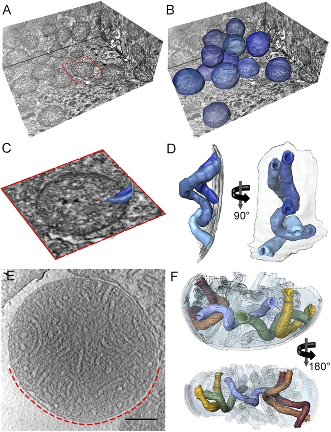
Fig. 4.
In situ morphology of mitochondria and mitochondrial cristae from P. tetraurelia. (A) Orthogonal slices through the tomographic volume of part of a P. tetraurelia cell generated by serial block face imaging with a focused ion-beam scanning electron microscope. (B) As A but with mitochondria segmented (blue). P. tetraurelia mitochondria are visible as discrete spheres. (C) Close-up view of the mitochondrion highlighted in A (red box) with segmented cristae (blue). (D) Surface representation of the cristae segmented in C. Transparent gray, inner membrane. See Movie S4. (E) Slice through an electron cryotomogram of an isolated plunge-frozen P. tetraurelia mitochondrion. (F) Surface representation of cristae in the region of the mitochondrion indicated by the dashed red line in E. Cristae of P. tetraurelia mitochondria are right-handed helical tubes of 40 nm diameter. The tubes are connected to the inner boundary membrane at both ends by circular cristae junctions. See Movie S5. The crista morphology in whole cells and in isolated mitochondria is indistinguishable. (E scale bar, 200 nm.) (C) Length of red box is ∼1.4 µm.

