Fig. 5.
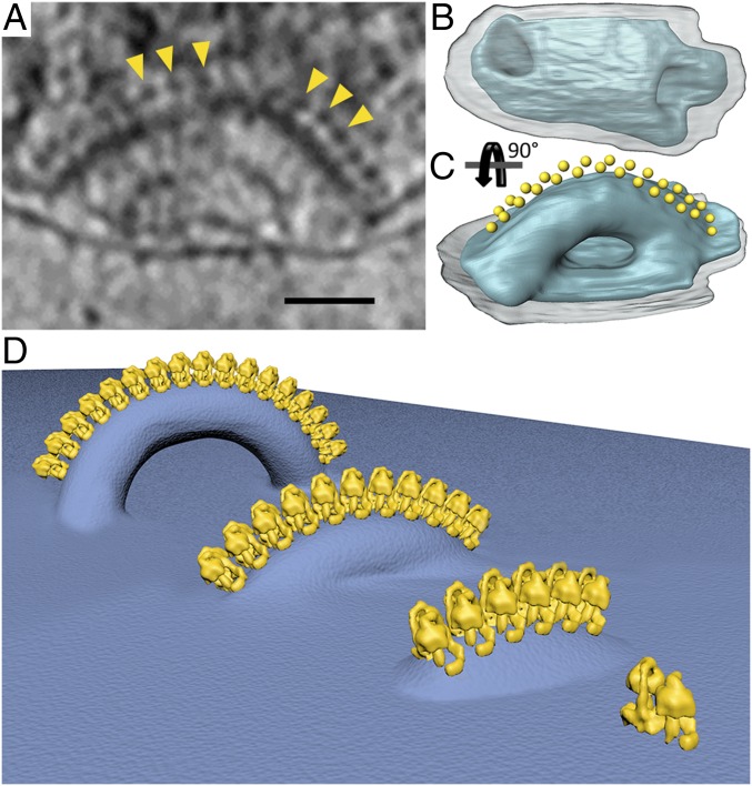
Formation of cristae. (A) Tomographic slice through a mitochondrion showing a short tubular crista decorated with ATP synthase dimers (yellow arrowheads). (B and C) Segmented surface-rendered representation of the crista shown in A (blue) with ATP synthase monomers represented by yellow spheres. The tubular crista forms a twisted arc with a right-handed helical array of interdigitated ATP synthases along its outer perimeter. Transparent gray, outer membrane. (D) Schematic of crista formation in P. tetraurelia. ATP synthase dimers associate into a helical array that deforms the membrane locally. The deformation becomes more pronounced as the length of the array increases, until the membrane below the row ruptures, forming a twisted tube with a crista junction at each end and a flat inner boundary membrane in between. (A scale bar, 50 nm.)

