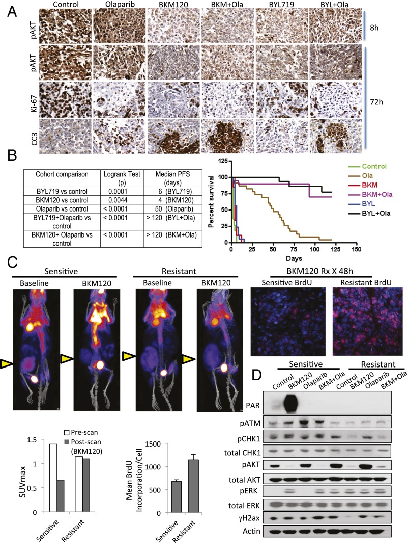
Fig. 4.
Response to PI3K and PARP inhibition in vivo. Six different primary tumors from K14-Cre BRCA1f/fp53f/f females were propagated in vivo through syngeneic transplantation. Recipient females were randomized to treatments with BKM120 (30 mg⋅kg⋅−1d−1 by mouth), BYL719 (30 mg⋅kg⋅−1⋅d−1 by mouth), Olaparib (50 mg⋅kg⋅−1d−1 i.p.), or their combination, and tumor volume was recorded every 2 d. Treatment endpoint was time to progression as defined by tumor volume doubling. (A) Two mice per treatment condition were killed after 8 and 72 h of drug treatments to assess biomarkers of response, as assessed with Ki67 and CC3 by IHC (magnification: 200×). These mice were given a last dose of drugs 2 h before killing. (B) Survival statistics (log-rank test) and Kaplan–Meier analysis of 132 recipient females randomized to treatments as indicated. (C) Resistance to PI3K inhibition. 18FDG-PET-CT scan at baseline and after 48 h of administration of the PI3K inhibitor NVP-BKM120 (3 doses, 30 mg/kg PO) in sensitive and BKM120+Olaparib resistant K14-Cre BRCA1f/fp53f/f tumor-bearing mice. The maximum standardized uptake value (SUVmax) of tumors before and after PI3K inhibitor administration is displayed in the bar graph. Tumors are marked with a yellow arrow. Two hours before killing, mice were injected with BrdU and given an additional dose of BKM120. Tumor sections were stained with anti-BrdU antibodies for immunofluoresence (red) (magnification: 200×) and mean fluorescence per cell of BrdU+ cells was measured using volocity software. (D) A cell line was derived from the clinically resistant tumor and its PI3K/PARP-sensitive parental tumor. Cells were treated in vitro with drugs as indicated for 16 h, lysed, and blotted with antibodies as indicated.

