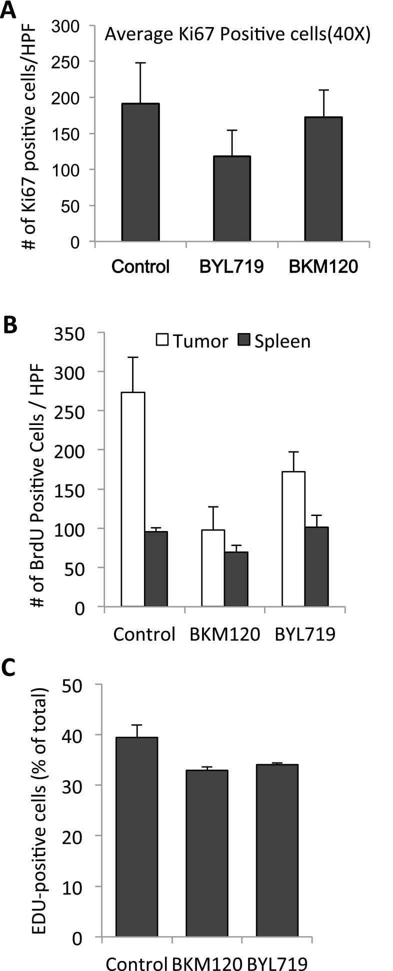
Fig. S1.
(A) Proliferative activity as assessed by Ki67 stain in tumors after 8 h of treatment with BKM120 (30 mg/kg) or BYL719 (30 mg/kg). In each cohort, three mice were treated 8 and 2 h before killing, three mice per cohort, tumors fixed and stained with anti-Ki67 antibodies, and five high power fields (HPF, 400×) counted. Differences between the three cohorts were not significant by t test (two-tailed, unpaired). (B) Percentage of tumor cells entering S phase upon PI3K inhibition. K14-Cre BRCA1f/fp53f/f tumor-bearing mice were prepared, randomized, treated, and labeled with BrdU as described in Fig. 2A. Bar graphs display the number of BrdU+ cells in tumors (solid) or spleens (light) per 40× field, representing the mean of 3–5 different tumors. Significance was P < 0.05 for the bars marked with an asterisk. (C) HCC1927 cells were treated as described in Fig. 2B. Bar graphs display the percentage of EdU+ cells, means of experimental triplicates for each condition.

