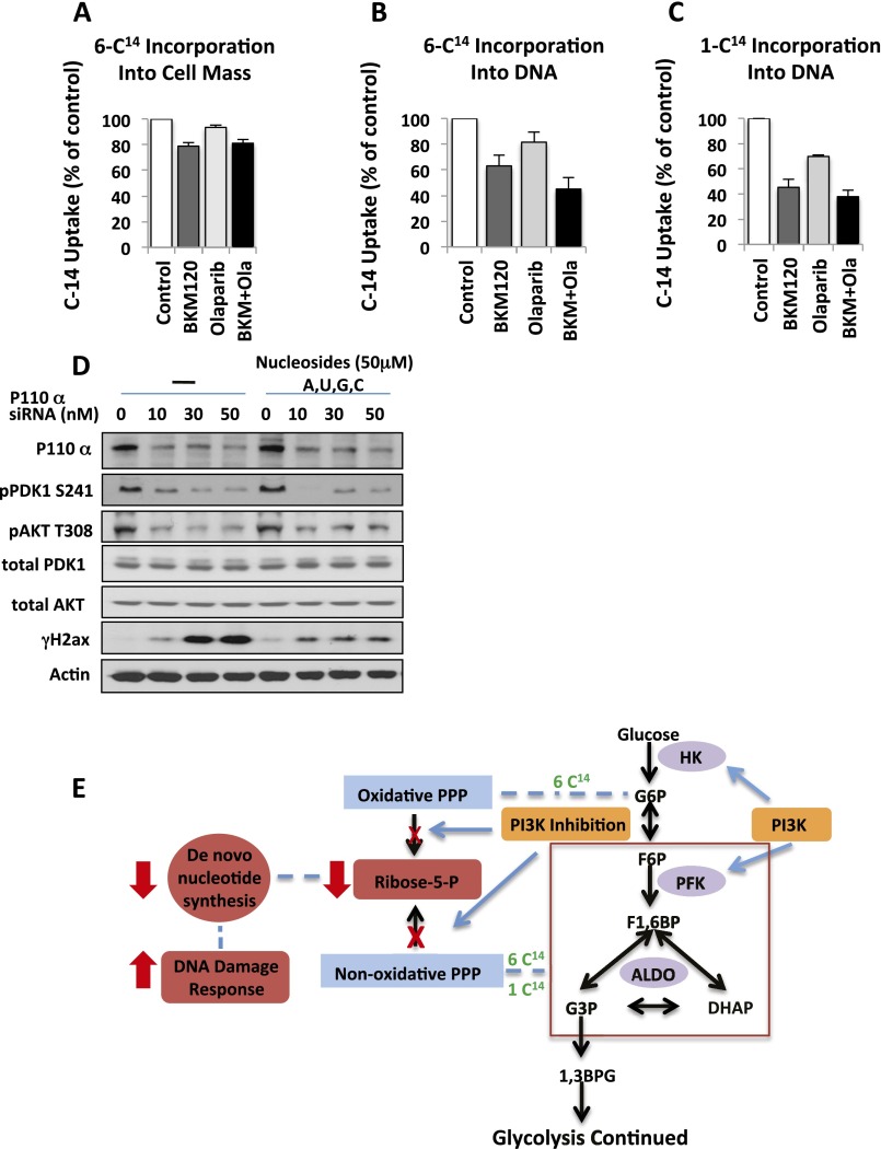
Fig. S3.
Carbon flux from glucose to Rib as determined by 14C-glucose–derived carbon into cell biomass (A) or DNA (B and C) in response to PI3K and PARP inhibition. HCC1937 cells were cultured in the presence of 14C6-glucose or 14C1-glucose, BKM120 (1 μM) or Olaparib (5 μM), or their combination for 3 h as indicated. Scintillation was counted for the entire cell lysate (A); genomic DNA was extracted and 14C measured (B and C). (D) Loss of PI3Kα leads to induction of H2AX phosphorylation (γH2AX) that can be rescued by nucleoside reconstitution. HCC1937 cells were depleted of PI3Kα by using siRNA for 48 h, and then treated with or without nucleosides for another 16 h. Cell lysates were subjected to immunoblotting with antibodies as indicated. (E) A radioactive label in the 6 position will measure flux through both oxidative and nonoxidative PPP, whereas a label in the 1 position will allow to measure flux through the nonoxidative PPP only.

