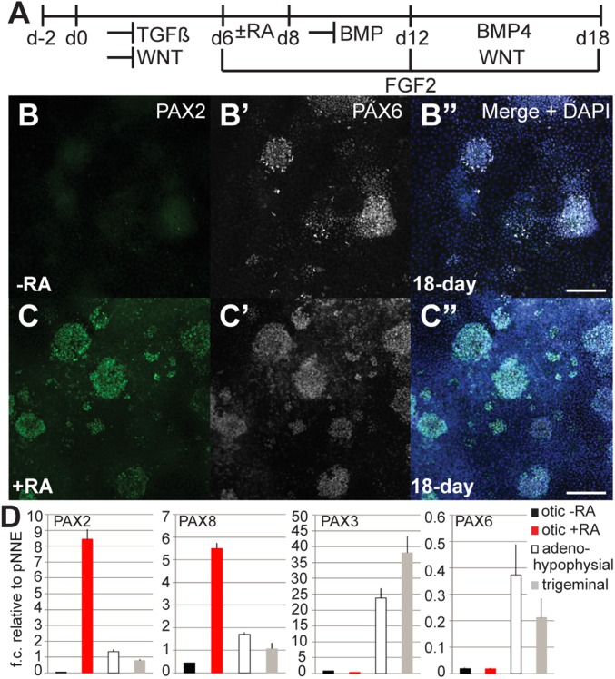
Fig. 4.
Posterior placode marker expression upon treatment with retinoic acid. (A) Monolayer culture conditions for otic induction. (B–B″) Immunocytochemistry showed no detectable expression of PAX2, but expression of PAX6 in cultures without retinoic acid (RA) treatment. (C–C″) Adding retinoic acid between days 6 and 8 resulted in expression of PAX2 detected at day 18 (14.1 ± 10.5%; n = 4). (D) qRT-PCR analysis of 18-d cultures for anterior and posterior placode markers. NNE cultures treated for otic induction with or without retinoic acid, adenohypophysial, and trigeminal placode induction. (Scale bars, 100 μm.)

